+1234-567-89

info@ivomedpk.com

+1234-567-89

info@ivomedpk.com

Home > Our Products > REVO-9

Electromedical
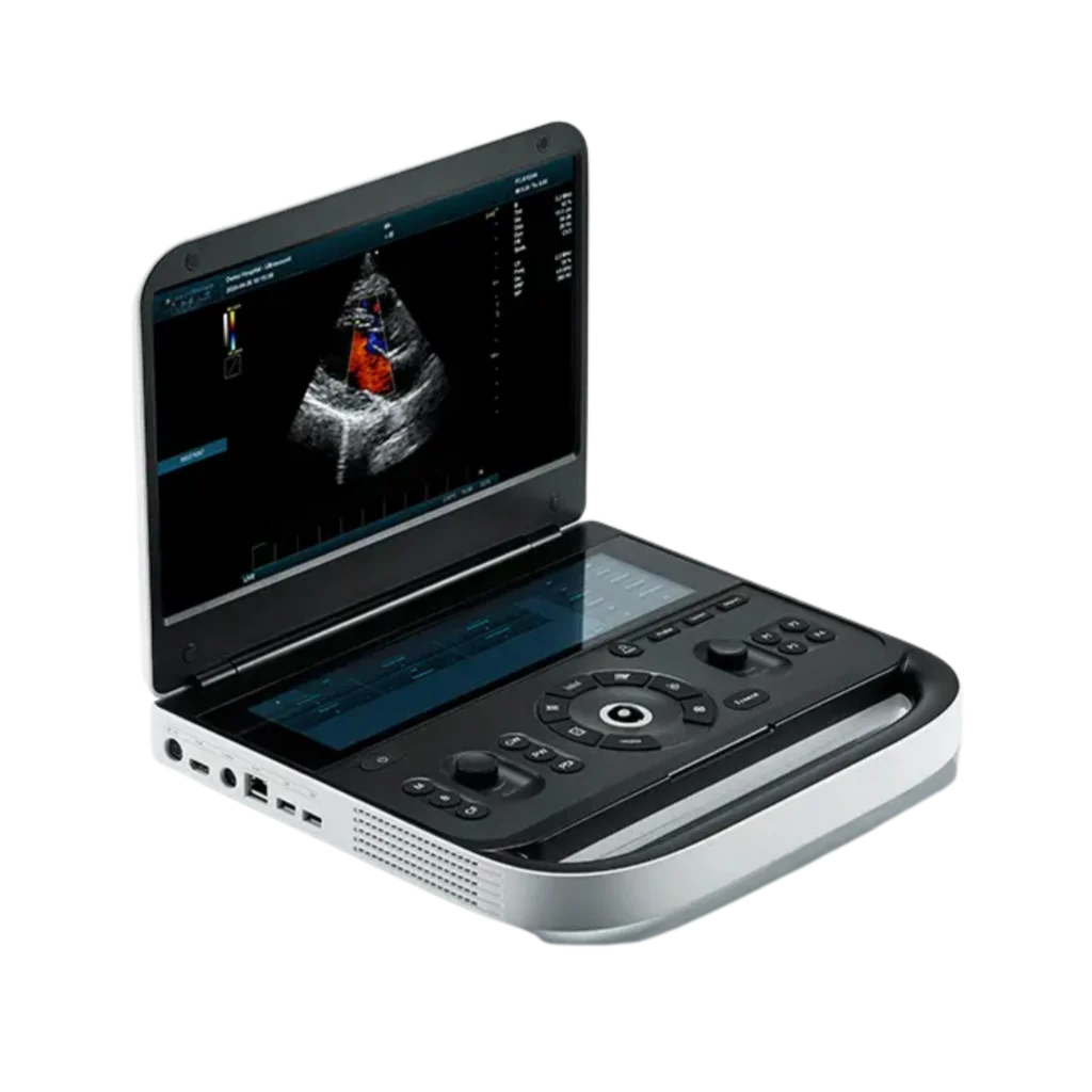
Premium Diagnostic Ultrasound System

REVO 9

Product Features

Nerves & Heart

Blood vessels

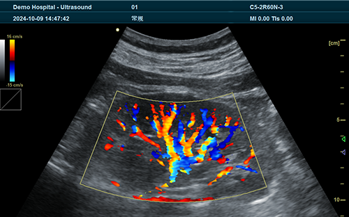
Urinary tract

Musculoskeletal system

Clinical Application in abdomen

Product Features

Nerves & Heart

Blood vessels

Musculoskeletal system

Urinary tract

Clinical Application in abdomen

Imaging

Intelligent Space Review Imaging

Color M Mode (MC)

It is used for cardiac examination. Color blood flow uses speed and vari ance colormaps to make colors super imposed on Mmode images.Color blood flow covers B-mode images and M-mode timeline.
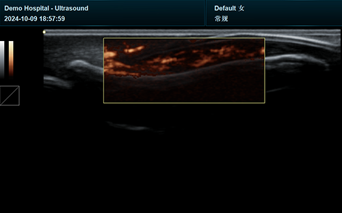
Tissue Doppler Imaging (TDI)

TDI enables the identification of spatial and temporal myocardial deformation distribution between different seg ments. By offering objective and reliable evaluation of myocardial move ment and function,
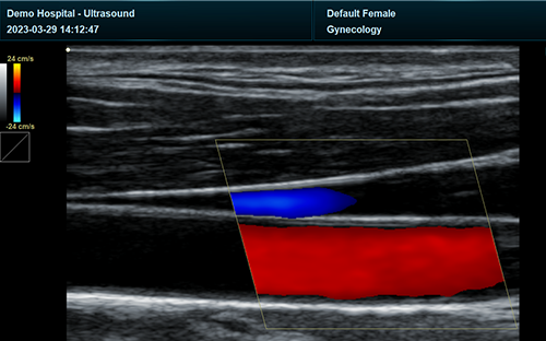
Anatomical 3M lmaging (AM)

AM is notrestricted by three orthogonal planes, thetechnology of artificially cuting volumedata in any direction and angle throughstraight lines, curves, polylines, and trajec-tories of any plane or angle.

PRODUCT DISPLAY

Excellent performance for you

By combining advanced adaptive precise transmission and reception control with efficient multi-channel parallel processing technology,
the machine’s computing speed is maximized while achieving the best image performance
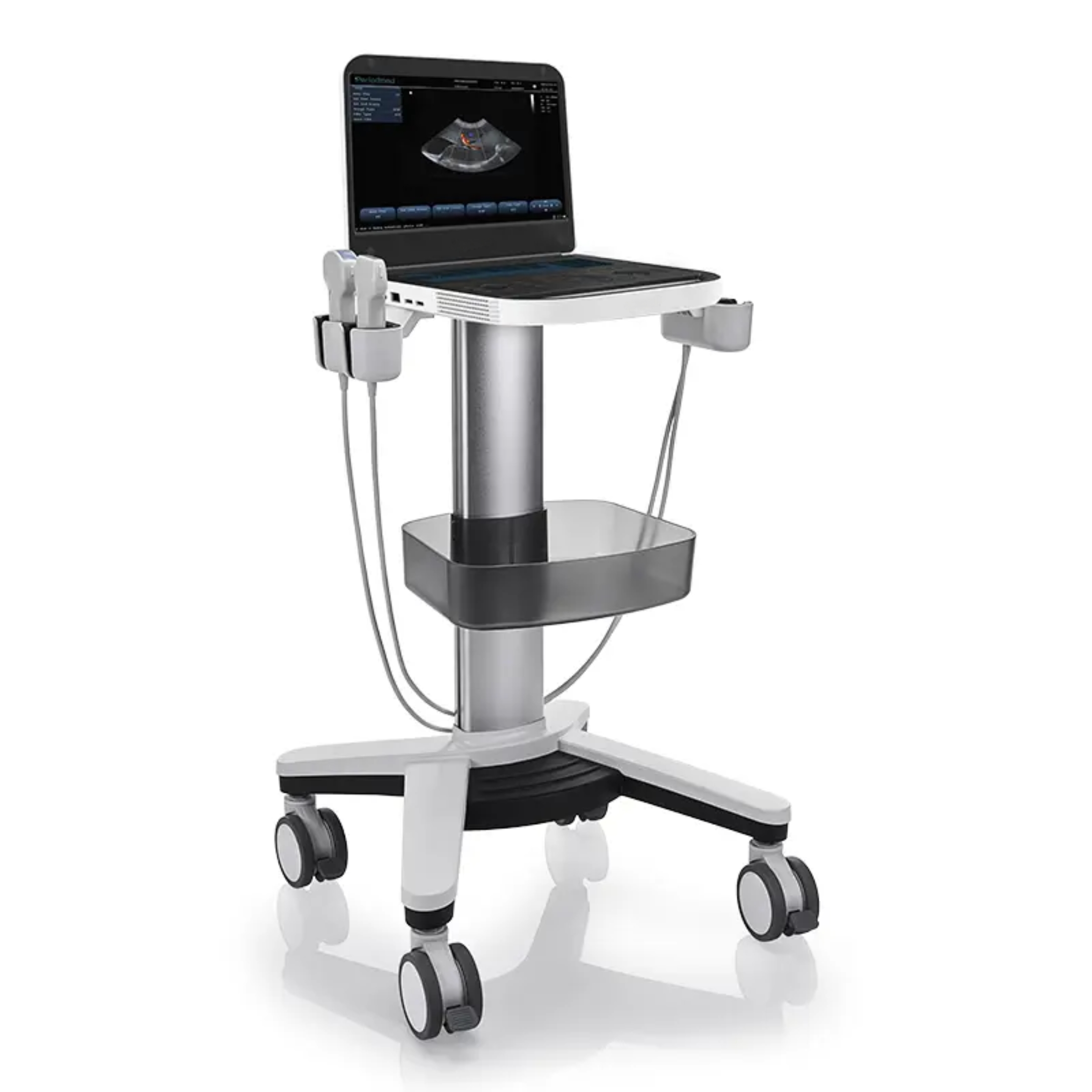
Technical Specification

Application Generic, Abdomen, Obstetrics, Gynecology, Vascular, Small Parts, Urology, MSK, Cardiology
Transducer Types Convex Array, Linear Array, Phased Array, Micro Convex Array, Trans-vaginal, Intracavity Probe
Operating Modes
  • B Mode, Dual B, Quad B, B/M Mode, CF Mode, M Mode, PW Mode
  • CW Mode, Power Doppler Imaging, Directional Power Doppler Imaging, Tissue Doppler Imaging, 3D/4D Mode, Wide View (Extend Field of View) (Optional), Picture in Picture
  • Trapezoid, Panorama, Color M Mode, Anatomy M Mode, Elastography (Optional), Needle Enhance Mode (Optional), Contrast Enhance Mode (Optional)
Imaging Technique & Function
  • Automatic Optimization, Spatial Compound Imaging, Speckle Reduction Imaging, Color Quantification, Fine Angle Steer, Tissue Harmonic Imaging (THI), Pulse Inversion Harmonic Imaging (iTHI)
  • Virtual Convex, Patient Information Database, Image Archive on Integrated Hard Drive, Real-time Automatic Doppler Calculation, OB Calculations, Fetal Trending, Multi-gestational Calculations
  • Hip Dysplasia Calculations, Gynecological Calculations, Vascular Calculations, Urological Calculations, Renal Calculations, Cardiac Calculations, On-board Electronic Documentation
  • PW Auto-trace, Privacy and Security, DICOM, Wide View, Auto IMT, External USB Printer Connection
Simultaneous Capability B/CF, B/PDI or DPDI, B/PW, C/M, B/M, B+CF or PDI or DPDI/PW or M
System Language Support English, Arabic, Chinese, French, German, Indonesian, Italian, Portuguese, Russian, Spanish
System Architecture
  • Physical Channels: 64
  • Frequency Range: 2.0 - 22.0 MHz
  • Beam Forming: 16
  • Elements: 64/128/160/192
  • Hard Drive: 256G
  • Memory: 8G
  • System Dynamic Range: 36 - 120db
Monitor
  • 15.6 Inch High Resolution LCD Monitor
  • Resolution: 1920*1080
  • 12.6 Inch Multifunctional Touch Screen
  • Magnetic Latch Closure
  • Built-in Speaker
  • Brightness and Contrast Adjustable
Various Universal Probe
  • C5-2R60N Convex Probe — Applications: Generic, Abdomen, Obstetrics, Gynecology, Urology; Frequencies: 2.5, 3.5, 4.0, 5.0, 6.0, H3.6, H4.0, H5.0, H6.0 MHz
  • C5-2R20N Microconvex Probe — Applications: Generic, Abdomen, Obstetrics, Gynecology, Urology; Frequencies: 2.5, 3.5, 4.0, 5.0, 6.0, H3.6, H4.0, H5.0, H6.0 MHz
  • P4-2L15SI Phased Array Probe — Applications: Generic, Abdomen, Cardiology; Frequencies: 2.0, 3.0, 4.0, H3.2, H3.6, H4.0 MHz
  • L12-5L40N Linear Probe — Applications: Small Parts, Vascular, Musculoskeletal, Superficial; Frequencies: 5.0, 7.5, 8.5, 10.0, H8.0, H9.0, H10.0, H12.0 MHz
  • C9-5R10N Transvaginal Probe — Applications: Obstetrics, Gynecology, Urology; Frequencies: 4.0, 5.0, 6.5, 8.0, 9.0, H7.0, H8.0, H9.0 MHz
  • L14-6L40H Linear Probe — Applications: Generic, Vascular, Small Part, Musculoskeletal; Frequencies: 7.5, 10.0, 12.0, 14.0, H10.0, H12.0, H14.0 MHz
  • C9-5 Volume Probe — Applications: Obstetrics, Abdomen, Urology; Frequencies: 5.0, 6.0, 9.0 MHz
  • L12-5L60N Trans-rectal Probe — Applications: Obstetrics, Gynecology, Urology; Frequencies: 5.0, 7.5, 8.5, 10.0, H8.0, H9.0, H10.0, H12.0 MHz

ADVANCED IMAGING TECHNOLOGIES

Our ProOpti™ system utilizes automatic structure detection algorithms to enhance image quality by sharpening edges and ensuring smooth, uniform tissue representation. This innovative feature enhances diagnostic accuracy by providing clearer and more detailed images.

By employing PrismX™ spatial composite imaging technology, our system enhances contrast, fine resolution, and spatial resolution. It optimizes tissue and pathological interface echo continuity while minimizing artifacts such as specular reflection, speckle, scattering, and attenuation, ensuring superior image clarity.

Our PureSight™ technology reduces speckle artifacts, improves smoothness along preferred edge directions, and maintains consistent local average gray levels. This results in clearer and more visually appealing images, enhancing diagnostic confidence.

PanoramaVision™ technology captures a series of two-dimensional sectional images, which are reconstructed into an ultra-wide sectional image with a
continuous field of view. This enables precise measurement of organ size and lesions, while providing a comprehensive view of the lesion's scope, internal echo, location, size, and adjacent tissues.

Get a Quote

Contact our team for pricing and availability of the REVO 9.